Strengthening early cancer detection through quality control
Muhammad Yaseen, a UICC Technical Fellow working at the Lady Reading Hospital in Pakistan, describes how a free, automated quality control software can make imaging equipment more reliable, and help detect cancers earlier.
When cancers are detected at stages 1 and 2, survival rates can exceed 80% for many common cancers. And when routine screening – one that is done at regular intervals for a target, at-risk population – identifies pre-cancerous lesions, the cancer can be prevented from developing.
Low- and middle-income countries, however, often face the challenge of late-stage diagnoses. This means that a cancer is diagnosed at stage III or IV, when the cancer is no longer localised and has spread to other parts of the body.
In these cases, cancer is more difficult to treat effectively, and is done so at a greater cost to the health system and to the patient – both financially, physically, and emotionally. In Pakistan, approximately 30% of people with cancer are diagnosed at stage III or stage IV.
Late-stage diagnosis is one of the main reasons for wide discrepancies in survival rates between high- and low-resource settings for certain cancers, particularly for cancers such as breast, cervical, and colorectal cancers. Initiatives to strengthen early detection, such as organised screening programmes, alongside efforts to strengthen follow-up for timely diagnosis, are key to reduce stage at diagnosis.
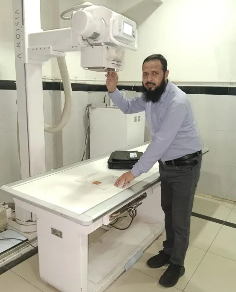
Muhammad Yaseen, Senior Medical Physicist & Radiation Protection Officer at Lady Reading Hospital in Peshawar, Pakistan, and a UICC Technical Fellow, is keen to emphasise, however, that even when organised screening programmes are available, the implementation of screening alone is not always sufficient. “It is not only a matter of whether imaging equipment exists, but whether it is working accurately enough to detect disease at a stage when treatment can still make a difference,” he explains.
With more than a decade of experience in diagnostic radiology, nuclear medicine, radiation protection, and quality assurance, Yaseen points to a range of challenges that can undermine the effectiveness of available screening capabilities.
“There are shortages of qualified medical physicists, quality assurance tools can be very expensive, and we often face infrequent maintenance checks. All this means that machines can produce images of insufficient quality for months before anyone identifies the fault,” he says.
The consequences for diagnosis can be significant. According to Yaseen, when equipment such as mammography or general x-ray machine is not properly calibrated or maintained, smaller lesions may not be detected, or scans may appear normal when they are not.
“In mammography, the ability to detect a lesion of half a millimetre or smaller can determine whether a cancer is found at an early or late stage,” he says. “When errors in machine performance go undetected, the images produced may lack the resolution needed to identify small lesions or calcifications.”
Quality control within reach with low-cost daily checks
Pakistan is not alone in this. Health systems in many LMICs face a similar situation, with insufficient availability of equipment, shortages of trained personnel and limited infrastructure.
According to Yaseen, maintaining imaging equipment to a standard that makes accurate diagnosis possible is a persistent challenge in many public hospitals, where quality control checks occur once or twice a year.
“If a mammography or general x-ray machine develops a fault between those checks, the problem can go undetected for months”, he says. “We performed annual quality control and biannual quality control after six months. But every day, it is impossible to perform quality control tests."
It was this gap that led Yaseen to apply for a UICC Technical Fellowship, supported in part by the Prevent Cancer Foundation, a UICC member organisation. He wanted to learn how to implement a system of routine, automated quality control suited to the constraints of a high-volume public hospital with a limited budget.
His fellowship took him to Hamad Medical Corporation in Qatar, a collaborating centre of the International Atomic Energy Agency (IAEA), where he trained in the IAEA's remote and automated quality control programme.
To test a machine’s accuracy, the system combines the use of ‘physical phantoms’ – objects designed to simulate tissue and test how accurately a machine reproduces a known image –with specialised software that automatically evaluates image quality and flags deviations from expected performance. The software is available free of charge.
"Instead of relying only on periodic manual testing, the system automatically evaluates image quality parameters and provides performance data," Yaseen explains. "This helps us identify problems early and ensures that imaging equipment can reliably produce high-quality images."
At Hamad Medical Corporation, Yaseen received hands-on training using the phantoms and, on completing his visit, was gifted a set phantom for mammography and general X-ray imaging to take back to Pakistan. Back at Lady Reading Hospital, he began running daily and weekly quality control checks, tests that had previously not been possible without costly equipment. He found errors in machines that had not been detected in the most recent annual check.
“Through this equipment, we can perform weekly quality control. Each phantom contains materials of different densities, simulating tissue and calcifications at defined thicknesses. A single image is enough to show whether the machine is resolving those structures at the required level. If the resolution falls below the threshold, the problem can be identified and the biomedical department notified the same day.”
Mammography machines are now tested before each session to confirm they can detect lesions down to a defined size, according to Yaseen. If the resolution is insufficient, the machine is taken out of service until it is corrected. "It automatically improves the diagnosis of the patients and will help in early cancer detection, basically in mammography," he says.
Yaseen’s fellowship also produced an exchange of knowledge in both directions. He shared with his hosts in Qatar two areas being studied at Lady Reading Hospital: measuring radiation dose received by family members who accompany people into the imaging room – common in Pakistan for cultural reasons – and estimating foetal dose when pregnant women undergo CT scanning. "I shared this idea with them, and they expressed their interest in collaborating with us to further explore these research areas."
He also introduced his hosts to a model used at Lady Reading Hospital that he calls a ‘learning club’. This is a regular, multidisciplinary gathering where staff from cardiology, radiology, and other departments share knowledge and experience across specialties.
Since returning to Pakistan, Yaseen has trained colleagues at Lady Reading Hospital and at neighbouring hospitals. He has also engaged directly with the Pakistan Nuclear Regulatory Authority, the body responsible for radiation safety oversight nationally, to support wider adoption of these practices at national level.
He has also submitted a formal report on the programme to his department head, who passed it to the hospital administration. His department is now in the process of purchasing additional equipment as a result. And Yaseen is working on producing the phantoms locally: “Hamad Medical Corporation shared with me the knowledge of how to make them, and I hope to share this methodology with the Pakistan Nuclear Regulatory Authority so that these phantoms can be distributed to hospitals across Pakistan, enabling other institutions to perform the same daily, weekly quality control checks.
Lady Reading Hospital is also in discussion to become the first hospital in Pakistan to formally collaborate with the IAEA and Hamad Medical Corporation in the remote quality monitoring programme, joining a small number of countries already sharing imaging data with the two organisations for independent evaluation.
"The main lesson from this fellowship for me was that high quality imaging is not only about technology," Yaseen says. "It's about consistency, quality assurance, patient safety, and teamwork. By strengthening quality assurance and quality control, we can improve our diagnostic accuracy, reduce unnecessary radiation exposure and support earlier cancer detection for patients in Pakistan.”
Last update
Tuesday 30 June 2026